
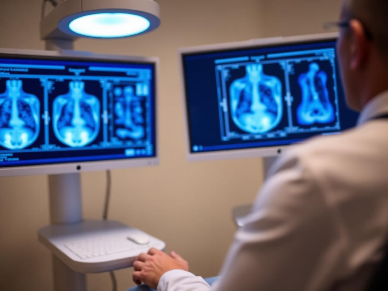
EXECUTIVE SUMMARY
- Dense breast tissue means the breast has more fibroglandular tissue than fatty tissue, which can make some findings harder to see on a mammogram.
- A breast density notification letter explains your density category and encourages you to talk with your physician about your personal screening plan.
- Supplemental screening dense breasts options may include 3D mammogram tomosynthesis, automated breast ultrasound or breast MRI screening, depending on your risk factors and physician guidance.
Dense breast tissue is a common finding on a mammogram report, and it can affect both your breast cancer risk profile and how clearly a mammogram can show small changes. If you receive a breast density notification letter in Glendale or elsewhere in Los Angeles County, it is meant to help you discuss whether supplemental screening for dense breasts, such as 3D mammogram tomosynthesis, automated breast ultrasound or breast MRI screening, may be appropriate with your physician.
Dense Breast Tissue what it means
Dense breast tissue means your mammogram shows a higher proportion of glandular and fibrous tissue compared with fatty tissue. This is not something you can reliably feel during a breast exam, and it is not the same as breast size or firmness. Breast density is determined by the radiologist who interprets your mammogram, based on how your breast tissue appears on the images.
On a mammogram, fatty tissue looks darker, while dense fibroglandular tissue looks whiter. Many breast findings, including some cancers, can also appear white. This overlap is one reason dense breast tissue can make mammogram interpretation more challenging. Your report may list density as one of several categories, ranging from mostly fatty to extremely dense.
A direct answer is: dense breast tissue is a mammogram description, not a diagnosis. It does not mean you have cancer, and it does not mean your mammogram was done incorrectly. It means your physician may consider your density along with age, family history, prior biopsies and other risk factors when planning breast screening.
At Lucent Medical Imaging in Glendale, breast imaging services are designed to support physician-directed screening and follow-up. You can learn more about dense breast screening options if your report notes heterogeneously dense or extremely dense breast tissue.
Dense tissue is seen only on imaging.
Density can change over time.
Your doctor interprets density with risk.
- Dense tissue is seen only on imaging.
- Density can change over time.
- Your doctor interprets density with risk.
Your Notification Letter why it arrives
A breast density notification letter is a required communication in many screening settings. Its purpose is to tell you that dense breast tissue was seen on your mammogram and that density may make cancer harder to detect on mammography alone. The letter is not an emergency notice and does not automatically mean you need another test.
The letter may use terms such as heterogeneously dense or extremely dense. Heterogeneously dense means there are many areas of dense tissue. Extremely dense means most of the breast tissue appears dense on the mammogram. These categories matter because they can affect image visibility and may slightly change how your overall risk is discussed.
What should you do after a breast density notification letter? You should read it with your mammogram result, then contact your ordering physician if you have questions. Your doctor can explain whether your mammogram was otherwise normal and whether supplemental screening dense breasts options fit your individual health history.
If you had screening in Glendale, Burbank, Pasadena or nearby Los Angeles County communities, your next step is usually a conversation with the clinician who ordered the exam. General information about breast imaging and screening can help you prepare questions before that visit.
Read the letter with your report.
Ask what your density category is.
Keep prior mammograms available.
- Read the letter with your report.
- Ask what your density category is.
- Keep prior mammograms available.
Questions before you book? We answer them.
Talk to our Glendale team about scheduling, preparation and what your plan covers, before you commit to an appointment.
Mammogram Visibility and dense tissue
Mammograms remain a central breast cancer screening tool, including for many people with dense breasts. However, dense tissue can hide or obscure small abnormalities because both dense tissue and some suspicious findings appear white. This is sometimes described as a masking effect, and it is one reason your physician may discuss additional imaging.
A 3D mammogram tomosynthesis exam takes multiple low-dose X-ray images from different angles and reconstructs thin image slices through the breast. These slices can help the radiologist evaluate overlapping tissue more clearly than standard two-dimensional mammography in many cases. It is still a mammogram, but it provides a more layered view of the breast.
Does a 3D mammogram solve dense breast tissue? A direct answer is: 3D mammogram tomosynthesis can improve tissue visualization, but it does not make dense tissue disappear and may not find every cancer. Your physician may still consider ultrasound or MRI depending on your density level and risk profile.
Lucent offers 3D mammogram tomosynthesis for screening and diagnostic breast imaging when ordered or appropriate. For many women, tomosynthesis is part of a practical screening plan, especially when prior mammograms show dense breast tissue.
Dense tissue can mask findings.
Tomosynthesis gives layered views.
Mammography remains important.
- Dense tissue can mask findings.
- Tomosynthesis gives layered views.
- Mammography remains important.
Supplemental Screening when considered
Supplemental screening dense breasts refers to imaging tests used in addition to mammography for some patients with dense tissue. These tests are not the same for everyone. Your doctor may consider your breast density, age, personal history, family history, genetic risk, prior biopsy results and comfort with possible follow-up testing.
The most common supplemental options include breast ultrasound, automated breast ultrasound and breast MRI screening. Each test looks at breast tissue in a different way. Ultrasound uses sound waves. MRI uses a magnetic field and radio waves, often with contrast when used for breast cancer screening. Mammography uses low-dose X-rays.
A clear answer is: you may need supplemental screening for dense breasts if your physician believes the potential benefit of finding an otherwise hidden cancer outweighs the chance of extra callbacks, additional images or biopsies. Dense tissue alone may not be the only factor in that decision.
Women in Glendale and nearby Los Angeles County areas can discuss women's health imaging with their ordering physician to understand which tests match their personal screening plan. The goal is not more testing for its own sake, but the right test for the right clinical question.
Supplemental tests add information.
Not everyone needs extra imaging.
Risk level guides the plan.
- Supplemental tests add information.
- Not everyone needs extra imaging.
- Risk level guides the plan.
Automated Breast Ultrasound how it helps
Automated breast ultrasound uses sound waves to create images of breast tissue without radiation. Unlike handheld ultrasound, an automated system captures broad, standardized images of the breast. This can be useful as supplemental screening dense breasts imaging because ultrasound can sometimes show findings that are difficult to see within dense tissue on a mammogram.
Sonocine automated whole breast ultrasound is one type of automated breast ultrasound used for dense breast screening. The exam is typically performed with you lying down while the system scans the breast in a planned pattern. A radiologist then reviews the images along with your clinical history and mammogram information when available.
What is automated breast ultrasound for dense breasts? It is an ultrasound-based screening exam that may be used in addition to mammography for patients with dense breast tissue. It does not replace a mammogram in most screening plans, because mammography can show calcifications and other findings ultrasound may not show as well.
Lucent offers Sonocine automated breast ultrasound as part of breast imaging services. Your physician can help decide whether automated breast ultrasound is appropriate after reviewing your mammogram report and overall risk factors.
Ultrasound uses sound waves.
It is often supplemental.
Mammograms are still needed.
- Ultrasound uses sound waves.
- It is often supplemental.
- Mammograms are still needed.
Breast MRI Screening for higher risk
Breast MRI screening is another supplemental option, most often considered for people at higher-than-average risk or for specific clinical indications. MRI does not use ionizing radiation. It creates detailed images using a strong magnetic field and radio waves, and breast screening MRI often uses contrast material to highlight areas of increased blood flow.
For dense breasts, MRI may be discussed when risk factors extend beyond density alone. Examples can include a strong family history, known genetic risk, prior chest radiation, certain biopsy findings or a prior breast cancer history. Your physician or breast specialist determines whether MRI is appropriate based on accepted screening guidance and your circumstances.
A direct answer is: breast MRI screening is not automatically required just because you have dense breast tissue. It may be recommended when dense breasts are combined with other risk factors or when your physician needs a more sensitive screening tool for your situation.
Lucent provides breast MRI screening using 3 Tesla MRI capability, including GE AIR Recon DL AI-assisted MRI technology where applicable. If MRI is ordered, staff will review general preparation steps, including safety screening for implanted devices or metal.
MRI is usually risk-based.
Contrast may be used.
Safety screening is required.
- MRI is usually risk-based.
- Contrast may be used.
- Safety screening is required.
Risk And Results put together
Breast density is one piece of your breast health picture. Your physician may combine density with personal history, family history, reproductive history, prior biopsies, genetic information and previous imaging results. This broader view helps determine whether routine mammography is enough or whether additional screening should be considered.
Your mammogram report may also include BI-RADS assessment language. This assessment is separate from breast density. Density describes the background tissue pattern. BI-RADS describes the radiologist's overall impression and recommendation, such as routine screening, additional imaging or follow-up. If the wording is unclear, ask your physician to review both parts with you.
What does a normal mammogram with dense breasts mean? It means no suspicious finding was reported on that mammogram, but dense tissue may reduce sensitivity. Your doctor may still discuss whether your personal risk supports supplemental screening dense breasts imaging, especially if you are in a higher-risk category.
Keeping prior studies available is helpful because radiologists compare current images with older ones to look for change. If you are new to Lucent, information on getting your imaging results can help you understand how reports are shared with you and your physician.
Density is not BI-RADS.
Normal results can still need context.
Comparison images are valuable.
- Density is not BI-RADS.
- Normal results can still need context.
- Comparison images are valuable.
Before Your Visit what to ask
Before your appointment, gather your recent mammogram report, breast density notification letter and any prior breast imaging records if they are available. If you have had biopsies, surgery or treatment, bring those details as well. This information helps your physician and imaging team understand what question the exam is meant to answer.
Useful questions include: What is my density category? Was my mammogram otherwise normal? Do I have risk factors beyond dense breast tissue? Would 3D mammogram tomosynthesis, automated breast ultrasound or breast MRI screening add useful information? How would an abnormal supplemental result be handled? These questions support a shared, physician-guided plan.
A practical answer is: prepare for dense breast screening by bringing prior images, knowing your family history and asking how each test would change your care. Imaging is most useful when the reason for the exam is clear and the results can be acted on appropriately.
For visit logistics at 647 W Broadway in Glendale, you can review before your visit information or contact Lucent Medical Imaging at (818) 679-2867. If your physician recommends breast-focused imaging, the mammography service overview explains available mammography options.
Bring reports and prior images.
Write down your questions.
Confirm the ordering plan.
- Bring reports and prior images.
- Write down your questions.
- Confirm the ordering plan.
Choosing Next Steps with your doctor
Dense breast tissue can feel confusing because it is common, usually not felt physically and often mentioned in a separate letter. The key is to treat the information as a prompt for discussion, not as a diagnosis. Your physician can explain how much density matters for you specifically.
Your next step may be routine annual screening, a 3D mammogram tomosynthesis approach, automated breast ultrasound, breast MRI screening or another plan based on your history. Some people need only standard follow-up. Others may benefit from supplemental screening dense breasts imaging. The decision should consider benefits, limitations and possible downstream testing.
A direct answer is: after you learn you have dense breasts, ask your physician whether your overall risk is average, intermediate or higher, and whether supplemental imaging is recommended. Do not assume that every dense breast report requires the same response.
Lucent Medical Imaging serves Glendale, Los Angeles County and nearby communities with mammography, ultrasound and MRI services used in physician-directed breast care. If your doctor has ordered imaging, you may use the request an appointment page to begin scheduling.
Density prompts a conversation.
Plans should be individualized.
Your physician guides screening.
- Density prompts a conversation.
- Plans should be individualized.
- Your physician guides screening.
Key takeaways
- Dense breast tissue is a mammogram finding.
- A notification letter is not a cancer diagnosis.
- 3D mammogram tomosynthesis may improve visibility.
- Automated breast ultrasound can add information.
- Breast MRI screening is usually risk-based.
Related at Lucent
Frequently asked questions
This article is general education, not medical advice. Talk with your physician about what is appropriate for your situation.
About the author

Lucent Clinical Team
Board-certified radiologists & imaging technologists
Every article is written and reviewed by the imaging team at our Glendale center, the same radiologists and technologists who read your study and walk you through the scanner room. We write what we find ourselves explaining at the front desk each week: what a scan feels like, what a report actually says, and what a bill is really made of.
Keep reading
What to expect during your first MRI scan
From check-in to results, here is exactly how an MRI appointment unfolds and what you can do to make it easier.
14 January 2026 · 9 min
MRI vs CT: how doctors choose between them
Both produce cross-sectional images, but they measure completely different things, which is why your physician orders one and not the other.
28 January 2026 · 9 min
Is a whole-body MRI right for you?
Preventive whole-body MRI is genuinely useful for some people and unnecessary for others. Here is how to tell the difference.
11 February 2026 · 8 min