
EXECUTIVE SUMMARY
- MRI and CT both create detailed internal images, but they use different technology and answer different questions.
- CT is generally faster and is often useful for bones, lungs, bleeding and urgent evaluation, while MRI is often preferred for soft tissue imaging.
- Your physician chooses based on your symptoms, medical history, exam findings and the information needed to guide care.
MRI vs CT scan is a common question when your physician orders imaging to evaluate pain, injury, infection or a possible disease. The best test depends on the body area, the clinical question, safety factors and how quickly images are needed.
Core differences *between the tests*
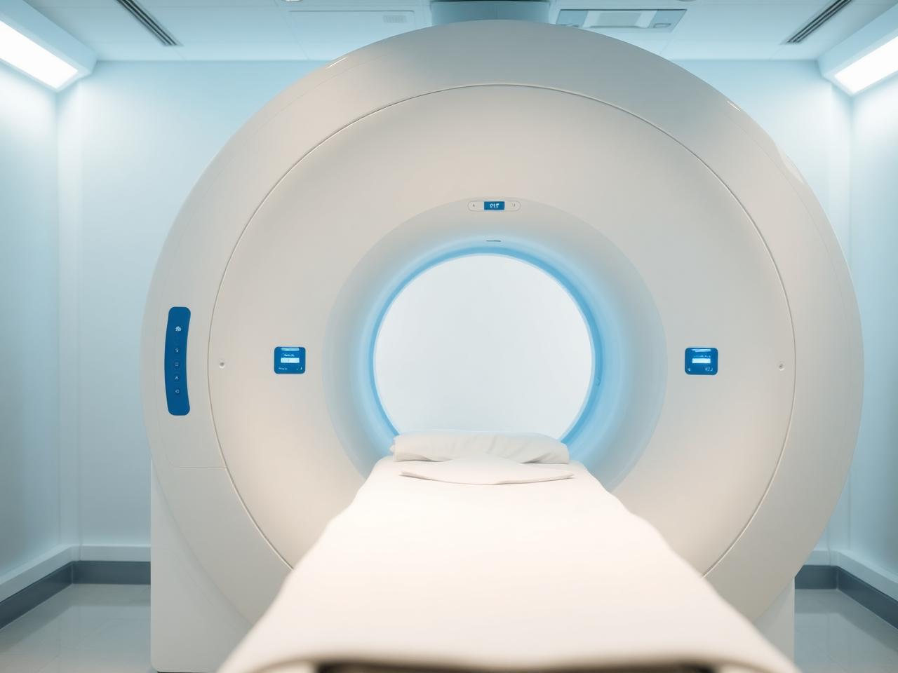
The difference between MRI and CT starts with how each test makes images. MRI uses a strong magnetic field and radio waves to show internal structures. CT uses X-rays taken from many angles, then a computer builds cross-sectional images. Both tests can be very detailed, but they highlight different tissues in different ways. In Glendale and across Los Angeles County, your physician may order either exam depending on whether the concern is in the brain, chest, abdomen, spine, joints or blood vessels.
A direct answer is this: MRI is often chosen when the main question involves soft tissue imaging, such as the brain, spinal cord, nerves, ligaments, muscles or certain organs. CT is often chosen when the question involves bone detail, lung disease, internal bleeding, kidney stones, trauma or fast evaluation. These are general patterns, not strict rules, because the right choice depends on your specific symptoms and the suspected diagnosis.
At Lucent Medical Imaging, MRI services include advanced MRI imaging, while CT services include diagnostic CT scan imaging. Your ordering physician decides which exam is medically appropriate, and the radiologist interprets the images in the context of the order. If you are unsure why a test was ordered, ask your physician what question the exam is meant to answer.
MRI does not use ionizing radiation.
CT uses X-ray technology.
Both may use contrast.
- MRI highlights many soft tissues well.
- CT is often faster.
- Your physician sets the imaging question.
How doctors decide *step by step*
Doctors usually begin with the clinical question. For example, the question may be whether there is a fracture, stroke, tumor, herniated disc, pneumonia, kidney stone or abdominal inflammation. The suspected problem narrows the choice. A CT may answer some questions quickly, while MRI may provide more tissue contrast for others. The exam is not chosen because one machine is universally better, but because one method is better matched to the question.
Another direct answer: doctors choose between MRI vs CT scan by weighing diagnostic value, speed, safety and patient factors. They consider your symptoms, physical exam, prior imaging, kidney function, implanted devices, pregnancy status when relevant, ability to lie still and whether contrast is needed. They also consider whether the result must be available quickly to guide urgent treatment.
Location matters too. A patient in Glendale with a sports injury may need joint or spine imaging, while someone with cough and abnormal chest findings may need chest CT. Nearby patients from Burbank, Pasadena, Eagle Rock and other Los Angeles County communities may be referred based on the same medical reasoning. For preparation details, your care team may also direct you to before your imaging visit.
The suspected diagnosis guides the exam.
Safety factors are reviewed first.
Prior imaging may change the plan.
- The question comes first.
- Speed can matter.
- Safety factors guide choices.
Questions before you book? We answer them.
Talk to our Glendale team about scheduling, preparation and what your plan covers, before you commit to an appointment.
When CT helps *for fast answers*
When is a CT better than MRI? CT is generally better when speed, bone detail, lung detail or detection of certain acute conditions is most important. CT is commonly used after trauma because it can quickly show fractures, internal bleeding or organ injury. It can also be useful for kidney stones, appendicitis, bowel obstruction and many chest concerns. In urgent settings, the speed of CT can be a major advantage.
CT is also strong for lung evaluation because air-filled structures are difficult to assess on many MRI exams. If your doctor is evaluating lung nodules, pneumonia complications, emphysema patterns or certain screening questions, CT may be the preferred tool. Lucent provides CT imaging for chest and lung concerns, including information on lung imaging focus areas for patients seeking general education.
Another common reason CT is selected is bone detail. CT can show complex fractures, joint alignment and small areas of calcification clearly. MRI may show bone marrow swelling and soft tissue injury, but CT often provides sharper detail of hard bone surfaces. Your physician may still choose MRI if the concern is ligament injury, disc disease, infection or a marrow process rather than a fracture line.
CT is often used after trauma.
CT can show lungs clearly.
CT provides strong bone detail.
- CT is useful for urgent questions.
- Lung problems often need CT.
- Bone detail is a CT strength.
When MRI helps *with soft tissues*
MRI is often preferred for soft tissue imaging because it can separate many tissue types without ionizing radiation. It is commonly used for the brain, spine, joints, pelvis, abdomen and breasts when the clinical question requires contrast between soft tissues. MRI can show cartilage, tendons, ligaments, nerves, discs, marrow and many organ details in ways that CT may not match for certain conditions.
A direct answer is this: MRI is often better than CT when the doctor needs detailed soft tissue imaging of the brain, spinal cord, nerves, muscles, ligaments or certain organs. For example, MRI may be used for suspected disc herniation, ligament tears, brain tumors, multiple sclerosis evaluation, pelvic soft tissue questions or certain liver and breast findings. The exact choice depends on the reason for the exam.
For neurologic symptoms, your physician may consider brain imaging services or a dedicated brain MRI exam, depending on the concern. Lucent Medical Imaging offers 3 Tesla MRI and GE AIR Recon DL AI-assisted MRI, which can support detailed MRI examinations when MRI is the appropriate test. Your physician and radiologist determine the protocol needed for the body part and clinical question.
MRI is useful for nerves and discs.
MRI can show ligament injuries.
MRI avoids ionizing radiation.
- MRI excels in many soft tissues.
- Brain and spine questions often use MRI.
- Exam protocols vary by diagnosis.
Radiation and dose *what to know*
CT radiation dose is an important part of the MRI vs CT scan discussion. CT uses ionizing radiation because it is based on X-rays. MRI does not use ionizing radiation. This does not mean CT should be avoided when it is medically needed. It means your physician weighs the expected benefit of the information against the radiation exposure, especially if you have had multiple prior scans or if the exam is for a younger patient.
A direct answer is this: low dose CT uses CT techniques designed to reduce radiation exposure while still producing images that are useful for a specific purpose. Low dose methods may be appropriate for selected exams, such as certain lung screening or follow-up studies, but not every CT can be performed at the lowest dose. The dose must still be adequate to answer the medical question.
Lucent offers low dose CT when appropriate for the ordered exam. If you are concerned about CT radiation dose, ask your ordering physician why CT is recommended and whether prior imaging could answer the question. You can also bring records from previous imaging so the radiology team knows what has already been done.
MRI has no ionizing radiation.
CT dose depends on the exam.
Low dose CT has specific uses.
- CT uses X-rays.
- Dose is matched to the question.
- Prior scans can be helpful.
Contrast decisions *and safety checks*
Both MRI and CT may be performed with or without contrast. Contrast is a substance used to make certain tissues, blood vessels or areas of inflammation easier to see. CT contrast is often iodine based. MRI contrast is usually gadolinium based. Whether contrast is needed depends on the reason for the exam, the body area and the information your physician needs.
Before contrast, the imaging team reviews safety information. You may be asked about kidney disease, allergies, prior contrast reactions, pregnancy status and current medications. For MRI, you may also be screened for pacemakers, implanted devices, metal fragments or other objects affected by the magnetic field. These questions help determine whether the exam can be done as ordered or whether adjustments are needed.
A direct answer is this: contrast does not make MRI or CT automatically better. It makes specific findings easier to see for specific questions. For example, contrast may help evaluate tumors, infection, inflammation, blood vessels or organ perfusion. Some exams are designed to be done without contrast, such as many kidney stone CT scans or some joint and spine MRI studies.
Contrast depends on the diagnosis.
Kidney history may be reviewed.
Implants matter for MRI safety.
- Contrast is not always needed.
- Screening questions protect safety.
- Protocols are tailored.
Comfort and timing *during the exam*
CT exams are usually quick, often with only a short time on the scanner table. MRI exams usually take longer because they collect different image sequences over time. During MRI, you must lie still inside or partly inside the magnet opening, and you will hear loud knocking or tapping sounds. Ear protection is typically provided. CT is quieter and more open, but both exams require you to hold still for clear images.
If you feel anxious in enclosed spaces, tell your physician and the imaging center before the appointment. Your doctor may choose the exam that best answers the question while also considering your ability to complete it. Some patients tolerate CT more easily because it is faster. Others complete MRI without difficulty when they know what to expect and receive clear instructions from the technologist.
Patients from Glendale and nearby Los Angeles neighborhoods often ask about scheduling, authorization and coverage before imaging. Insurance rules can affect timing because some plans require authorization before MRI or CT. Lucent provides general help through insurance information for patients, but your benefits and medical necessity rules come from your insurer and ordering physician.
CT is usually shorter.
MRI is louder.
Stillness improves image quality.
- Tell the team about claustrophobia.
- Authorization may affect timing.
- Clear instructions help.
Body area examples *common choices*
For the head, the choice depends on the question. CT may be used when a doctor needs a fast look for bleeding, skull fracture or certain urgent changes. MRI may be used when the question involves brain tissue, nerves, tumors, inflammation or subtle changes that are not as clear on CT. This is why two people with headaches may receive different imaging orders.
For the spine and joints, MRI is often used for discs, nerves, cartilage, ligaments and tendons. CT may be chosen for fracture detail, surgical planning or areas where bone anatomy is the main question. A knee injury, back pain or shoulder problem may require different imaging depending on whether the physician suspects a soft tissue injury, a bone injury or both.
For the chest and abdomen, CT is frequently used for lungs, stones, bowel problems, trauma and many abdominal emergencies. MRI may be used for selected liver, pelvic, cardiac, breast or soft tissue questions. A physician may also use ultrasound, mammography or X-ray when those tests are more appropriate. The best imaging test is the one that answers the specific question with appropriate safety and clarity.
Head imaging depends on urgency.
Joints often need soft tissue detail.
Chest concerns often use CT.
- Symptoms shape the order.
- Different body parts favor different tools.
- No single scan fits all.
Your next steps *after the order*
Once your physician orders imaging, review the exam name, body part and whether contrast is requested. Ask what diagnosis is being evaluated and how the result may affect your care plan. If you have had prior MRI, CT, ultrasound or X-ray studies, bring the reports or images if requested. Prior exams can help the radiologist compare changes over time.
If your physician orders MRI, you can learn more about Lucent's 3 Tesla MRI technology. If CT is ordered for lung screening or a chest concern, your doctor may refer to a specific protocol such as lung screening CT. Use the exact order from your physician when scheduling, since the protocol must match the clinical question.
After the scan, a radiologist reviews the images and sends a report to your referring physician. Your physician explains what the findings mean for you, because imaging results must be interpreted with your symptoms, exam and medical history. If you have urgent symptoms or worsening symptoms, contact your physician or seek emergency care rather than waiting for a routine imaging appointment.
Know the ordered body part.
Bring prior imaging if asked.
Your physician explains results.
- Confirm contrast instructions.
- Use the exact order.
- Follow up with your doctor.
Key takeaways
- MRI is often best for soft tissue detail.
- CT is often best for speed, lungs and bone detail.
- CT uses radiation, MRI does not.
- Low dose CT may fit selected exams.
- Your physician chooses based on the medical question.
Frequently asked questions
This article is general education, not medical advice. Talk with your physician about what is appropriate for your situation.
About the author

Lucent Clinical Team
Board-certified radiologists & imaging technologists
Every article is written and reviewed by the imaging team at our Glendale center, the same radiologists and technologists who read your study and walk you through the scanner room. We write what we find ourselves explaining at the front desk each week: what a scan feels like, what a report actually says, and what a bill is really made of.
Keep reading
What to expect during your first MRI scan
From check-in to results, here is exactly how an MRI appointment unfolds and what you can do to make it easier.
14 January 2026 · 9 min
Is a whole-body MRI right for you?
Preventive whole-body MRI is genuinely useful for some people and unnecessary for others. Here is how to tell the difference.
11 February 2026 · 8 min
Understanding your imaging bill and insurance coverage
Why the same scan can cost wildly different amounts, and the questions to ask before you book.
25 February 2026 · 9 min