
How Sonocine automated breast ultrasound works
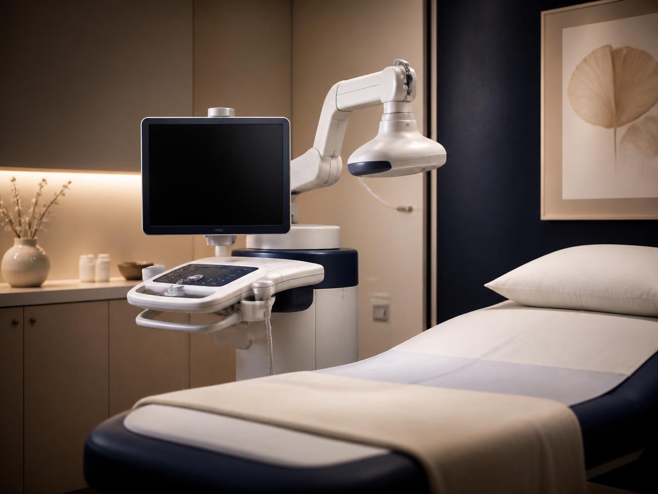
Sonocine automated breast ultrasound uses a motorized transducer to scan the entire breast in a systematic pattern, capturing a complete image set for a radiologist to review. It supplements mammography for women with dense breast tissue, with no radiation and no compression.
This page explains the technology. Ready to book?
See our Sonocine service pageWhen dense tissue hides things.
Dense fibroglandular tissue appears white on a mammogram, and so do many tumors. When there is a lot of dense tissue, a small cancer can sit inside that whiteness and be genuinely difficult to see, even for an experienced breast radiologist reading a perfectly performed study.
Density is common: roughly 40% of women have dense breasts. It is not a disease and it is not something you did, but it does reduce mammographic sensitivity, and it is an independent risk factor for breast cancer. California law requires that patients be notified of their breast density, and if yours is dense, supplemental screening is worth a calm conversation rather than an anxious one.
Handheld vs automated ultrasound.
Four steps, start to finish.
Guided pass
A motorized transducer is guided across the breast in a defined, repeatable pattern.
Thin-slice capture
Hundreds of thin-slice images are captured automatically as the transducer travels.
Set assembly
The slices are assembled into a continuous, reviewable image set covering the whole breast.
Radiologist review
A breast-imaging radiologist reviews the full set alongside your mammogram.
What supplemental screening can and can’t do.
Supplemental ultrasound increases cancer detection in dense tissue, but it also increases the chance of finding something benign that needs a follow-up image or a biopsy. That trade-off is real and worth knowing before you book.
Sonocine supplements mammography; it does not replace it. If we find something, we explain it clearly, in person, and coordinate the next step with you rather than leaving you to interpret a report alone.
Breast imaging at Lucent.
3D Mammogram
Thin-slice tomosynthesis, the foundation of breast screening.
Read moreDense Breast Screening
What breast density means and how we screen for it.
Read moreBreast MRI
High-sensitivity imaging for high-risk screening and problem solving.
Read moreSonocine service page
Who it is for, what to expect and how to book your screening.
Read more