
Breast imaging, matched to your risk
Screening, dense-tissue supplements and problem-solving studies in one center. We help you and your physician choose the study that answers your specific question, not just the one you were expecting.
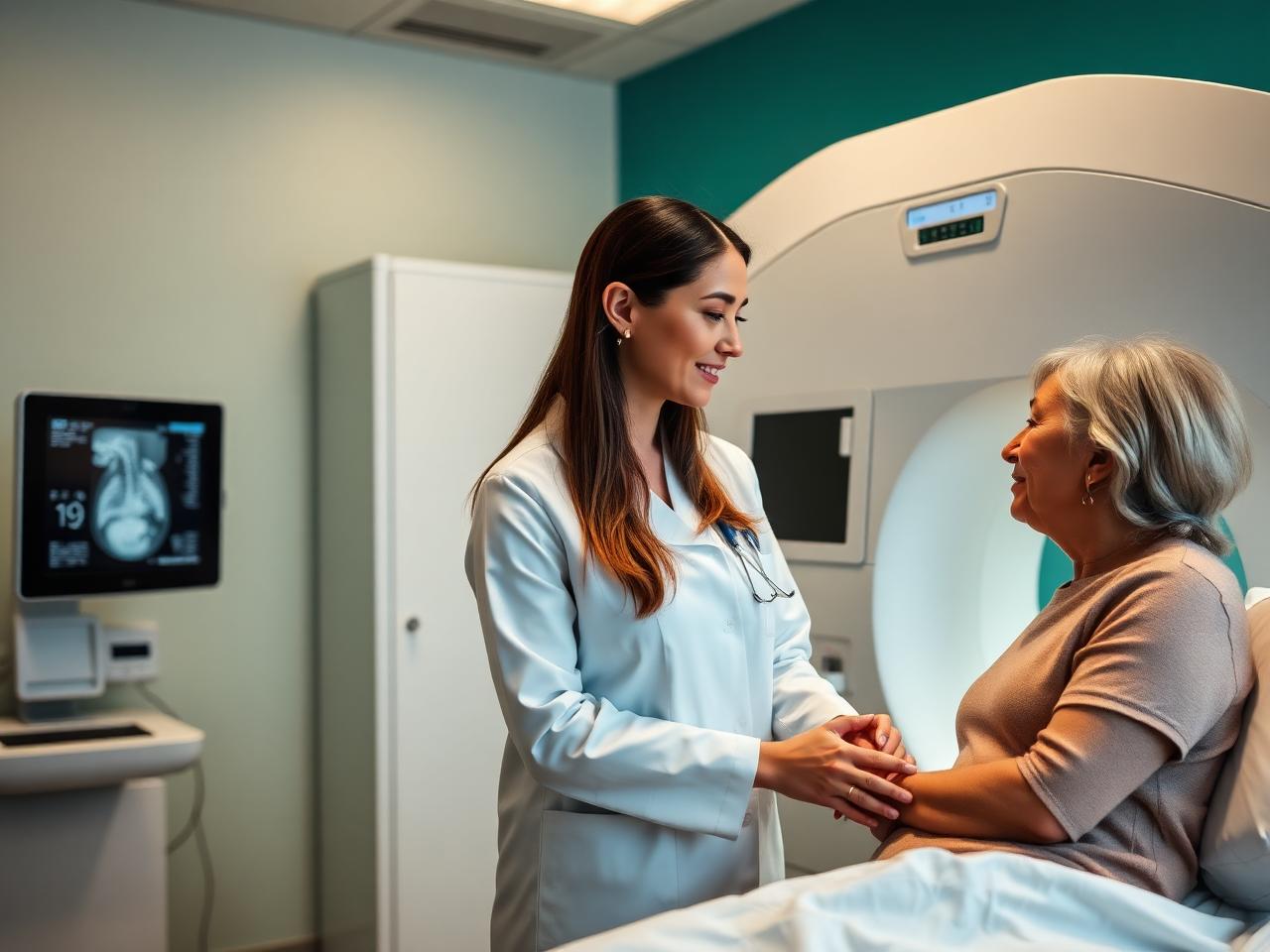
- 3D tomosynthesis screeningMAMMOGRAPHY
- Dense tissue supplementSONOCINE
- High-risk surveillanceBREAST MRI
- Reports to your physician24-48 HRS
Questions and conditions this imaging answers
Imaging does not replace your physician's evaluation, it gives them the detail needed to act with confidence.
Reasons patients come to us
- Annual screening mammogram
- A palpable lump or focal breast pain
- Dense breast tissue noted on a prior report
- Nipple discharge or skin changes
- Callback after an abnormal screening
What imaging can show
- Masses, cysts and architectural distortion
- Microcalcification patterns
- Extent of a known finding before treatment
- Changes compared with prior studies
- Findings in tissue mammography alone can hide
Which scan answers which question
Different questions call for different studies. Use this as a guide, then confirm the right exam with your physician or our scheduling team.
| Your question | Study most often used | Why that study |
|---|---|---|
| I am due for routine screening. | 3D mammogram (tomosynthesis) | Layer-by-layer images reduce the overlap that causes both missed findings and false callbacks. |
| My report says I have dense breast tissue. | Sonocine automated whole-breast ultrasound | Adds an ultrasound layer where dense tissue can mask a finding on mammography. |
| I feel a lump. | Diagnostic mammogram plus targeted breast ultrasound | Ultrasound distinguishes a simple cyst from a solid mass in the specific area of concern. |
| I am considered high risk. | Breast MRI with contrast | Highest sensitivity for early cancers; used alongside, not instead of, mammography. |
| I have a new diagnosis and need staging. | Breast MRI | Shows the extent of disease and screens the opposite breast before treatment planning. |
Final exam selection depends on your clinical history, prior imaging and insurance authorization.
What breast imaging can and cannot do
No single breast study detects every cancer. Supplemental imaging finds cancers mammography can miss, and it also finds benign changes that lead to extra visits or biopsies.
- Supplemental ultrasound and MRI add to screening mammography; they do not replace it.
- Some findings require short-interval follow-up or biopsy to resolve.
- Contrast-enhanced MRI requires a kidney function check for some patients.
Frequently asked questions
Do I need a referral for a screening mammogram?
Most insurance plans require a physician order for screening mammography. Call us at (818) 679-2867 and our team will confirm what your plan requires.
Is 3D mammography better than 2D?
Tomosynthesis images the breast in thin layers, which improves detection in many patients and reduces the chance of being called back for overlapping tissue.
Why was I told to add an ultrasound?
Dense breast tissue and cancer both appear white on a mammogram. Ultrasound provides a second look at tissue where a finding could be hidden.
How soon will I get results?
Reports typically reach your referring physician within 24 to 48 hours for routine studies.