
Neurological answers at 3 Tesla clarity
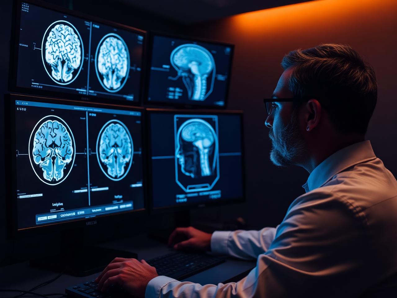
Headache, dizziness, memory change and neurological symptoms deserve high-resolution imaging. Our 3 Tesla MRI resolves small structures that lower field strength can blur.
- Double the field strength of 1.5T3 TESLA MRI
- MS, memory and seizure protocolsSUBSPECIALTY
- No radiation with MRISAFETY
- Reports to your physician24-48 HRS
Questions and conditions this imaging answers
Imaging does not replace your physician's evaluation, it gives them the detail needed to act with confidence.
Symptoms that lead here
- New, severe or changing headaches
- Memory or cognitive change
- Numbness, weakness or vision change
- Dizziness, vertigo or hearing loss
- Seizure workup or known epilepsy
What imaging can show
- Demyelinating lesions consistent with MS
- Tumors, cysts and pituitary lesions
- Old or evolving stroke and small-vessel change
- Aneurysms and vascular malformations with MRA
- Volume loss patterns relevant to memory change
Which scan answers which question
Different questions call for different studies. Use this as a guide, then confirm the right exam with your physician or our scheduling team.
| Your question | Study most often used | Why that study |
|---|---|---|
| I have new or worsening headaches. | Brain MRI, with or without contrast | Best soft-tissue detail for tumors, inflammation and structural causes. |
| My neurologist suspects MS. | Brain MRI with MS protocol, often with spine MRI | Thin-slice FLAIR sequences show the lesion pattern and location that support diagnosis. |
| I need to rule out an aneurysm. | MR angiography (MRA) of the head | Maps the intracranial arteries without radiation and, in many cases, without contrast. |
| I hit my head and need urgent assessment. | Head CT | Fast and excellent for acute bleeding and skull fracture, the right first study in trauma. |
| I have neck or arm pain with numbness. | Cervical spine MRI | Shows disc herniation and nerve root compression that cause radiating symptoms. |
Final exam selection depends on your clinical history, prior imaging and insurance authorization.
Before your brain MRI
MRI uses a strong magnet, so implanted devices and metal history matter. Tell us about any of the following when you schedule.
- Pacemakers, defibrillators, neurostimulators or cochlear implants
- Aneurysm clips, metal fragments or prior eye injury from metal
- Pregnancy, kidney disease or a prior contrast reaction
- Claustrophobia, tell us in advance so we can plan your comfort
Frequently asked questions
Will I need contrast for a brain MRI?
It depends on the question being asked. Contrast is often used for tumor, infection and MS activity assessment, and is frequently unnecessary for routine structural imaging.
How long does a brain MRI take?
Most brain studies take 30 to 45 minutes depending on the protocol and whether contrast is used.
What is the advantage of 3 Tesla?
Higher field strength delivers more signal, which can be used for finer detail or shorter scan times, useful for small structures like the pituitary and inner ear.
Can I have an MRI if I am claustrophobic?
Yes. Tell us ahead of time. Many patients do well with our wide-bore system, headphones and a technologist in constant contact; your physician may also prescribe medication.