
Pinpoint what is causing the pain
Shoulders, knees, hips, spine and small joints. High-resolution MRI shows the ligament, cartilage and tendon detail that guides physical therapy, injection or surgery.
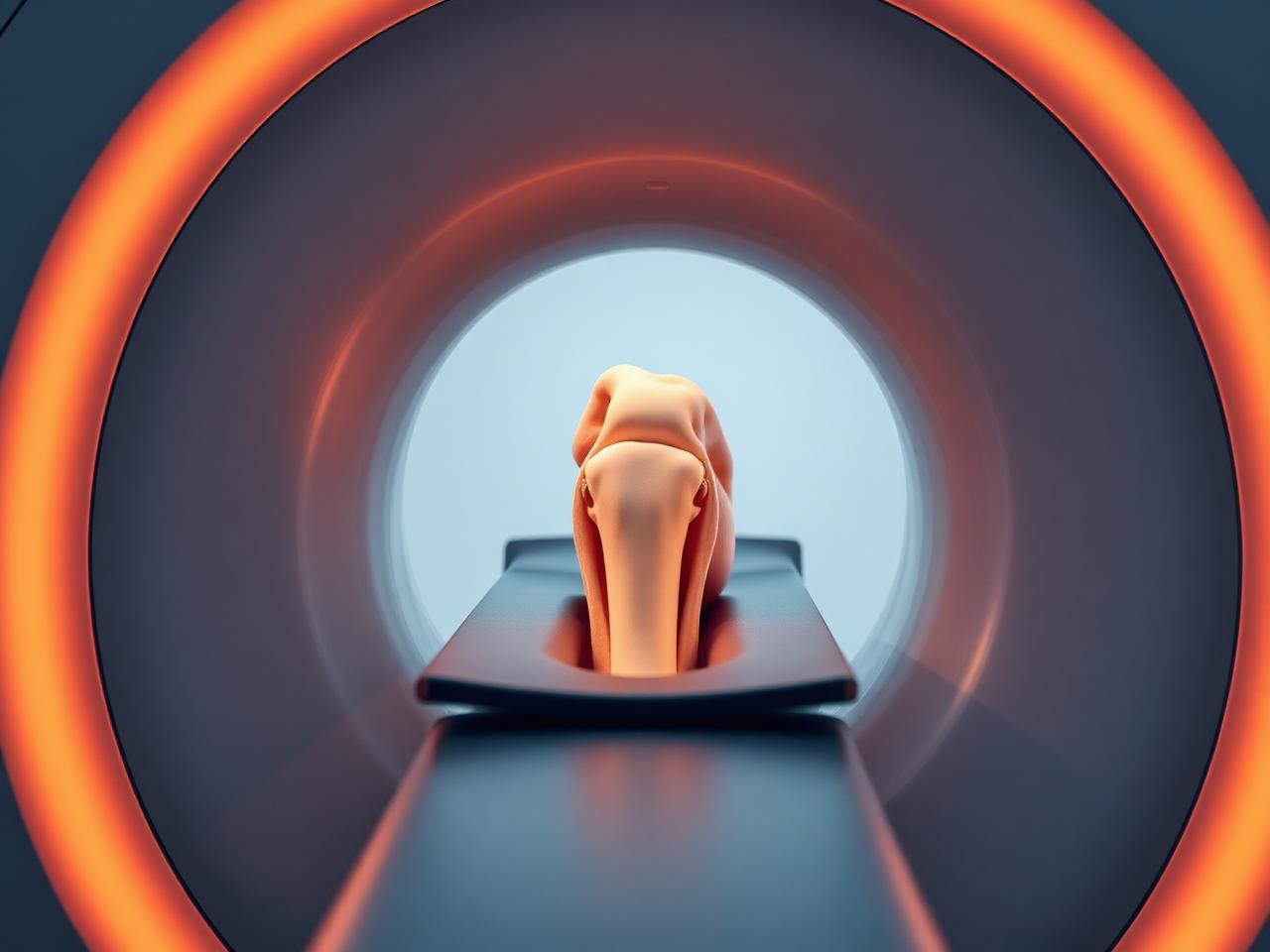
- Ligament, cartilage and tendon detail3T MRI
- Same-day fracture imagingX-RAY
- Dynamic soft-tissue assessmentULTRASOUND
- Reports to your physician24-48 HRS
Questions and conditions this imaging answers
Imaging does not replace your physician's evaluation, it gives them the detail needed to act with confidence.
Reasons patients come to us
- Sports injury or a joint that gave way
- Back or neck pain radiating into a limb
- Persistent shoulder, knee or hip pain
- Suspected fracture after a fall
- Pre-surgical planning or post-operative follow-up
What imaging can show
- Meniscal, labral and rotator cuff tears
- Ligament injury and cartilage loss
- Disc herniation and nerve root compression
- Stress fractures and bone marrow edema
- Tendinopathy, bursitis and fluid collections
Which scan answers which question
Different questions call for different studies. Use this as a guide, then confirm the right exam with your physician or our scheduling team.
| Your question | Study most often used | Why that study |
|---|---|---|
| I injured my knee or shoulder. | Joint MRI | Only MRI shows meniscus, labrum, cartilage and ligaments in full detail. |
| I may have broken something. | X-ray first | Fast and definitive for most fractures; MRI is added when X-ray is negative but suspicion remains. |
| My back pain radiates down my leg. | Lumbar spine MRI | Demonstrates disc herniation, stenosis and the nerve root actually being compressed. |
| I have a lump or tendon pain near the surface. | Musculoskeletal ultrasound | Real-time imaging shows tendons and soft-tissue masses while the joint moves. |
Final exam selection depends on your clinical history, prior imaging and insurance authorization.
Imaging findings are read alongside your symptoms
Degenerative changes are common on MRI in people with no pain at all. Your physician interprets the report in the context of your examination.
- Not every finding on a scan explains your symptoms.
- Metal implants may require a modified protocol, tell us at scheduling.
- Some joint studies use an injected contrast arthrogram; your physician will specify.
Frequently asked questions
How long does a joint MRI take?
Most single-joint studies take 20 to 40 minutes. Only the joint being studied goes into the center of the magnet where possible.
Do I need contrast?
Most routine joint MRIs do not. Contrast is used for infection, tumor and some post-surgical or labral questions.
Can I have an MRI with hardware from surgery?
Usually yes. Most orthopedic implants are MRI-safe; we adjust the protocol to reduce metal artifact. Bring your implant card if you have one.
Do I need a referral?
Insurance-covered MSK imaging requires a physician order. Call (818) 679-2867 and we will confirm what your plan needs.